Lung cancer: AI “eyes” improve diagnosis and disease prediction

According to a new study, artificial intelligence algorithms can identify the location of immune cells in histopathological samples of lung cancer, with significant implications for predicting disease course and therapy response
Thanks to artificial intelligence, it is possible to enhance our capacity for tumors analysis and description in histopathological slides, also by recognizing the position of subsets of immune cells with high precision. The result was achieved by training an image recognition algorithm using data from a cutting-edge technology called “imaging mass cytometry” – available in Humanitas and a few other research centers across Europe – capable of tracing up to 40 different immune response markers in the tumor microenvironment.
The algorithm obtained from this learning process has been consistently able to deduce the location of immune cells in the normal slides used in anatomic pathology. The achievement could change the work of pathologists in the near future, improving our capacity for diagnosis and prediction of therapy response.
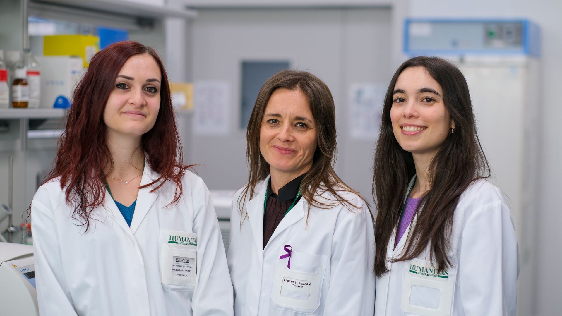
The study, which focused on the most common form of lung cancer but could be replicated and extended to other types of cancer, was published in Cancer Research by a team of researchers and doctors from Humanitas Research Hospital, led by Federica Marchesi, researcher in the Cellular Immunology Laboratory and associate professor at the University of Milan.
The research was possible thanks to the collaboration with specialists from the Thoracic Surgery and Anatomic Pathology Unit of Humanitas Research Hospital, as well as the Multiscale Imaging Unit, and was supported by the Italian Alliance Against Cancer and by AIRC Foundation for Cancer Research.
The Eyes of Artificial Intelligence
We are increasingly accustomed to artificial intelligence algorithms able to recognize images of any kind with great precision. These algorithms promise to revolutionize the field of anatomic pathology as much as many other fields of human activity: anatomic pathology relies on microscope observations of tissue samples to identify pathological conditions, such as tumors, by observing visible shape and structure – including the number and position of cells, and the arrangement of surrounding connective tissue and fibrosis.
“One of the most interesting aspects of AI algorithms is that, at least in some cases, they are able to deduce from visible profiles and shapes even sub-visual characteristics, that is, properties that can be inferred from what is seen but not in an obvious way,” explain Rebecca Polidori and Marika Viatore, lead authors of the study along with Alessandra Rigamonti. “A good example is facial recognition algorithms that understand the personality profile of the subject from the micro-expressions of their face.”
Humanitas researchers decided to exploit this sub-visual analysis capability to improve the assesment of lung cancer histological samples, especially the most widespread form, the “non-small cell” type. These tissue samples are stained with a technique commonly used called hematoxylin-eosin, which generically highlights the shape of cell nuclei, the cytoplasm surrounding them and the fluid-rich spaces that constitute the tissue itself. Only the experienced eye of the pathologist is able, based on these very simple elements, to recognize the presence of a tumor and identify its type. However, the question arises: what capabilities does an appropriately trained artificial intelligence algorithm hold?
Training AI with Data from One of the Most Sophisticated Experimental Imaging Tools
To train the AI to identify features in samples beyond those highlighted by hematoxylin-eosin staining, researchers led by Federica Marchesi provided the algorithm with highly detailed tissue information that can be extracted using imaging mass cytometry, an advanced imaging tool present in a few institutes across Europe: “The instrument allows us to trace with great precision the geography of the tumor microenvironment because it allows us to recognize up to 40 different proteins and molecules and therefore to distinguish the position of many types of cells – tumor and non-tumor, especially immune cells,” explains Federica Marchesi. “This is crucial, as recent research indicates that the relative positioning of immune cells reveals much about a tumor’s molecular characteristics, aggressiveness, and therapy response.”
First of all, the analysis conducted on lung cancer samples confirmed the key role of immunity in cancer: patients with better prognosis were those in which immune cells had managed to infiltrate the tumor more extensively, also thanks to the different arrangement of tumor cells. Knowing the position of immune cells thanks to this advanced imaging technique, Humanitas researchers were then able to train AI algorithms to infer it based on the tissue structure, even in samples stained only with hematoxylin-eosin, in which immune cells are not selectively traced.
“The study demonstrates that AI can be a powerful tool to enhance the analysis of histological slides and suggests a way to sustainably bring into the clinic the knowledge produced by increasingly widespread multidimensional analysis tools in research labs, like imaging mass cytometry,” concludes Federica Marchesi. “Indeed, these are technologies capable of identifying many markers but they are still expensive and slow to use in a diagnostic context: AI, properly trained with data produced by these tools, could help us make the most of classic diagnostic techniques, which are more readily accessible, by ‘seeing’ beyond what is strictly visible.”